Transwell实验 |
您所在的位置:网站首页 › transwell insert › Transwell实验 |
Transwell实验
|
Transwell检测细胞迁移步骤 1. 饥饿处理细胞 2. 铺胶:将基质胶适当稀释,铺在上层小室中,聚合过夜。 3. 铺下室培养基:含20%FBS的培养基。 4. 制备细胞悬液:胰酶消化细胞,制备细胞悬液。 5. 铺细胞:将上层小室放入孔板中,细胞计数后将细胞铺在上层小室中。 6. 继续培养:细胞培养箱中培养,通常24或48h。 7. 擦去上层细胞:用棉签擦去上层小室中的细胞和基质胶。 8. 固定细胞:4%多聚甲醛固定20min。 9. 染色与拍照:结晶紫或吉姆萨染色后拍照。 实验注意事项 基质胶注意事项 1. matrigel matrix成固态,液面水平。 2. 整个matrigel matrix只能分装一次,在分装前,要先放在4℃冰箱(最好先把matrigel matrix插在冰上,然后放入4℃冰箱)一段时间使其液化,且分装用的枪头,EP管等都要提前-20℃预冷(因为基质胶在10℃以上即会发生成胶凝固导致基质胶报废),整个分装过程要在冰上进行。 3. 实验人员的手心不要接触基质胶,如手持装有胶的EP管的上方,防止提问使基质胶成胶凝固。 4. 普通的matrigel matrix的浓度是8-12mg/ml,高浓度的是18-22mg/ml。 SCI写作范文及模板 1. PMID:31131011 Methods Transwell migration assay and transwell invasion assay The transwell migration assay and transwell invasion assay were conducted with a Corning Inc. transwell chamber. In the migration assay, 2 × 10⁴ cells suspended in 100 μl serum-free DMEM were seeded in the upper compartment of the chamber and 800 μl DMEM with 10% FBS were added to the lower compartment of the chamber. The NC mimic or miR-107 mimic were added at the same time and the cells were incubated for another 24 h. After that, the cells were fixed with 4% paraformaldehyde for 30 min and stained with 0.1% crystal violet. The non-migrating cells in the upper chamber were removed carefully using a cotton swab. The migrated cells were cells on the lower surface were photographed with an Olympus IX70 inverted microscope in five randomly selected visual fields and the migrated cells were quantified using Image-Pro Plus 6.0 software. Each assay was performed at least three times. For the invasion assay, the upper compartment was precoated with 100 μl of Matrigel. All other processes were the same as for the transwell migration assay. Figure legend 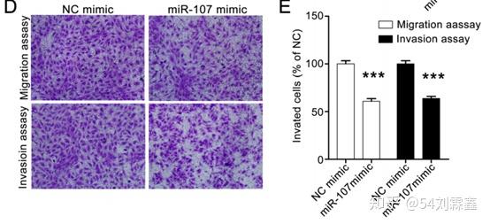 d and e MiR-107 supresses the vertical migration and invasion abilities of SW620 cells. The effect of miR-107 on vertical migration and invasion was assessed with transwell migration assay and tranwell invasion assay, respectively. Representative images (100× magnification) are shown in D and the quantitative data are shown in E. The data were analyzed with GraphPad Prism 5.0, and are presented as means ± SEM, n = 3. *** p < 0.001 compared with the control group. Results MiR-107 obviously inhibited the vertical migration and invasion of SW620 cells (Fig. 2d and e). 2. PMID:32699205 Methods Transwell assay Transwell assays were conducted using a transwell chamber coated with or without Matrigel. The 10% FBS medium was added in the lower chamber, and colon cancer cells (4 × 10⁴ /well) in serum-free medium were added to the upper chamber. After incubation at 37 °C with 5% CO2 for 24 h, the cells on the lower surface were fixed with methanol and stained with 0.1% crystal violet. 3. PMID: 32963602 Methods Transwell migration assay for assessing cell invasion ability. The Transwell migration assay was performed using the 24‑well Transwell with gelatin‑coated polycarbonate membrane filter. Firstly, cells were plated on a 6‑well plate at a density of 1x10⁶ cells/well and cultured until they reached 90‑95% confluency. 1, 2, 4, 8 µM/ml PTX solution was then added into the each well for 48 h of incubation. Subsequently, the treated cells were detached from the plates using 0.5 ml trypsin and then seeded in the upper chambers which precoated with 500 ng/ml Matrigel solution (BD Biosciences) of the Transwell chamber (~1x10⁴ cells/well), while DMEM supplemented with 10% FBS was added into the lower cham‑ bers. The cells were then incubated in the chambers for 24 h at 37˚C. Finally, the chambers were stained with 0.1% crystal violet at room temperature for 30 min, and imaged using a light microscope at x200 magnification. Figure legend 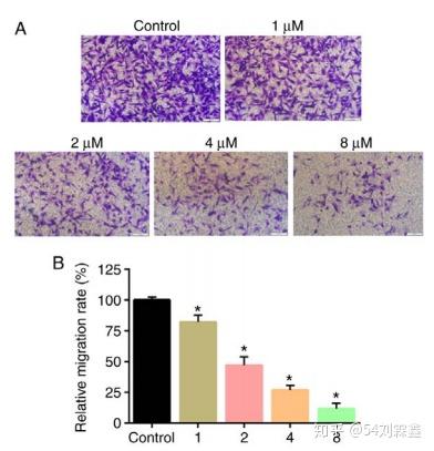 Inhibitory effect of PTX on invasion of CD133+ U251 glioma cells. (A) Transwell assays of CD133+ U251 glioma cells following treatment with PTX. The intensity of the crystal violet dye showed that cell groups treated with PTX showed reduced cell invasion compared with the control group, in which the PTX groups presented the lowest levels of cell invasion. (B) The relative migration rates of the observed groups. Data are presented as the mean ± standard deviation of three independent repeat experiments and analyzed using one‑way ANOVA followed by Tukey's post hoc test. Results Transwell assays were used to evaluate in vitro cell migration. Transwell assay results revealed that PTX treatment significantly attenuated the migratory ability of the cells compared with controls (Fig. 5A). The differences in the colony count and the migratory ability was found to be statistically significant. 4. PMID: 27941907 Methods Migration and Invasion assays. Cell invasion was determined using a transwell matrigel invasion assay in 24-well Transwell units (Costar). Matrigel diluted with the precooled serum-free Ham’s F-12K (50 μl) was added to the upper chamber of the Transwell and incubated at 37 °C for 30 minutes. The siRNAs or NC was prepared as described above. Following incubation for 20 minutes, 100 μl of the siRNA mixture was mixed to 100 μl serum-free Ham’s F-12K containing 1× 10⁵ cells and then transferred to Matrigel coated top chambers. The lower chambers were filled with 500 μl Ham’s F-12K supplemented with 10% FBS. After incubation at 37 °C for 24 hours, the non-invading cells were removed with a cotton swab. The inserts were removed from the top chambers, washed with PBS, fixed and stained with Giemsa. The invaded cells were counted in five random fields under a light microscope. The procedure for cell migration is similar to that of invasion, except that matrigel was not added. Figure legend Knockdown of Annexin A1 by siRNA inhibits AGS cell invasion. Invasion assay was conducted with Annexin A1 siRNA or NC transfected AGS cells using 24-well Transwell chambers. (a) Representative images of invaded cells in NC and siRNA transfected cells. (b) Cell invasion was assessed by counting the number of AGS cells that invaded through the transwell insert by light microscopy on 3 independent membranes, then normalized against the NC treated cells to determine the relative ratio. The procedures for cell invasion are similar to that of migration except that matrigel was added to the upper chamber of the transwell. 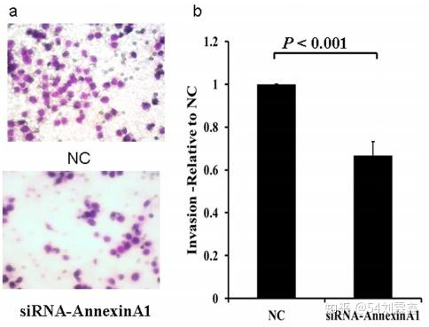 5. PMID: 28507454 Methods Migration and invasion assays Migration assays were conducted using 24-well Boyden chambers containing inserts (8 μm pores; BD Biosciences, USA). Invasion assays were performed similarly using matrigel-coated inserts (BD Bioscience, USA). The lower chamber was filled with medium containing 10% serum, whereas the top chamber contained 1 × 10⁵ HeLa cells or 5 × 10⁴ SiHa cells suspended in medium without serum. The plates were incubated at 37 °C in 5% CO2 for 24h. After migration, non-migrated cells remaining on the top of the inserts were removed with cotton swabs, whereas the cells that had migrated to the underside of the membrane was fixed with paraformaldehyde and stained with 1% crystal violet. Photos were captured with a fluorescence microscope (Olympus IX71, Japan). Migrated cells on each insert were counted in six randomly selected high-power fields (under a 20× objective lens) and quantified using the ImageJ software. Scratch assays were conducted on confuent cells and seeded in six-well plates. After the cells had adhered to the plates, the wells were gently scratched with straight lines using 20 μl sticks. When cells migrated into the wounds, the area decreased, during which cells were imaged every 12 for 48 h under a microscope (OLYMPUS IX71, Japan). For each experimental condition, the areas devoid of cells in six unique felds (under a 20× objective lens) were measured using ImageJ software. |
【本文地址】